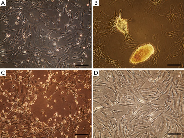
Figure 1.
Morphology of enriched cells. Isolated cells displayed heterogeneous morphologies as typical fibroblastic (A), spontaneous nodule-like structures (B) and spherical cytoplasm with extended processes (C). In control cultures, cells mostly possessed fibroblastic morphology (D). Scale bar =40 µm.

