Figure 4.
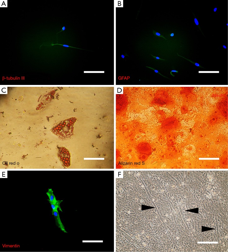
Differentiation potential assay of isolated SKPs. After 7 days of induced neuronal and glial differentiation, (A) β-III tubulin is expressed in a subpopulation of cells that are morphological consistent with neurons (Scale bar =30 µm). (B) Other subpopulation of differentiated SKPs expressed the glial marker GFAP (Scale bar =30 µm). (C) Lipid vacuoles (Red spot) were visualized after staining cells with oil red O which stains triglyceride and neutral lipid (Scale bar =20 µm). (D) Calcium deposition (Orange spot) was shown by alizarin red S staining after culturing cells in ostegenic medium for 15 days (Scale bar =20 µm). (E) Myogenin, marker of newly born myoblasts, was shown by immunocytochemistry in differentiated SKPs (Scale bar =20 µm). In addition, (F) myotube-like structures (arrow heads) were clearly visible in high confluent cultures of SKPs (Scale bar =40 µm).

