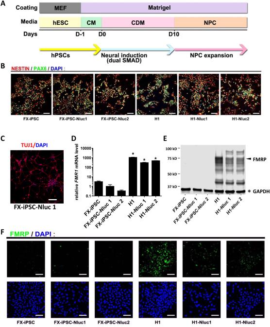
Figure 3. Neural differentiation and characterization of NPCs derived from reporter and parental lines.
(A) Schematic diagram of neural differentiation. (B) Differentiated NPCs are positive for neural progenitor markers, NESTIN and PAX6, as assessed by immunofluorescence (scale bar = 100 μm). (C) NPCs derived from the FXS reporter line were expanded for 23 passages and then differentiated into neurons (Tuj1+, red; scale bar = 50 μm). (D) Relative FMR1 mRNA levels assessed by quantitative RT-PCR. The mean expression of FX-iPSC-Nluc1 cells was set as 1 (Y axis in log scale; n=3; mean ± SEM; * indicates significant p value < 0.05). (E) A representative western blot result showing FMRP (arrow) and GAPDH (asterisk). (F) FMRP in reporter and parental NPCs detected using immunofluorescence and confocal imaging (scale bar = 100 μm).

