
Figure 5.
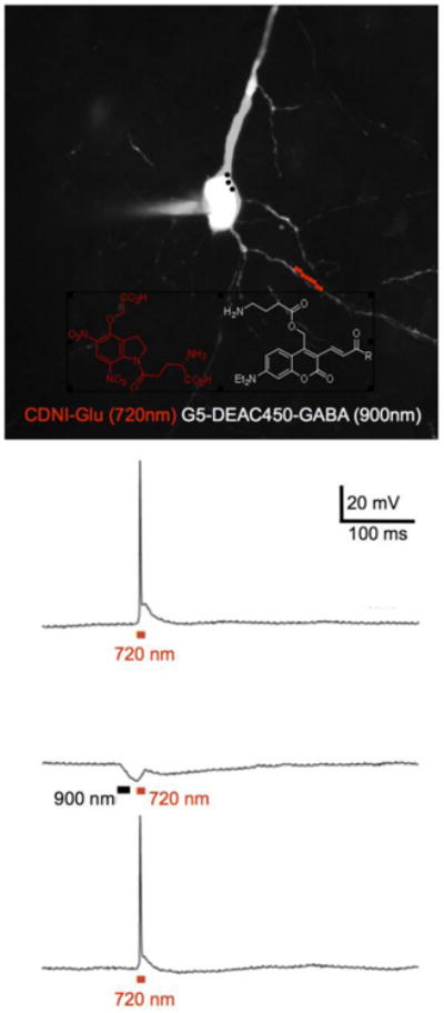
Chromatically orthogonal two-photon uncaging of glutamate and GABA. Example of a fluorescent image of a CA1 pyramidal neuron filled with Alexa594 via a patch pipette taken with excitation at 1070 nm. The red and black dots indicate the experimental protocol for irradiation at 720 nm along a dendrite (red dots) and 900 nm around the soma (black dots). CDNI-Glu and G5-DEAC450-GABA probes were bath applied at 1 mM and 0.6 mM, respectively. Uncaging at 720 nm (10 × 1 ms, 50 mW) fired an action potential (top trace), which could be blocked (middle trace) by prior uncaging at 900 nm (3 × 3ms, 50 mW). Such block was found to be reversible (lower trace).