Fig. 1.
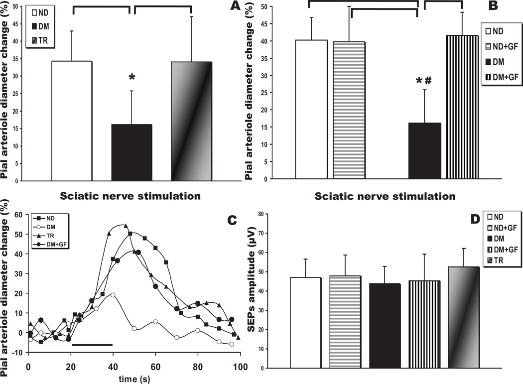
Neurovascular coupling impairment in 4 month diabetic (T1DM) rats. (A) Sciatic nerve stimulation (SNS)-evoked pial arteriolar responses in type 1 diabetes mellitus rats (DM, n = 6) show a ~45% decrease in the peak diameter change compared to non-diabetic controls (ND, n = 11) or animals subjected to pancreatic islet transplantation (TR, n = 7) (*, P < 0.05). (B) SNS-evoked responses before and after topical suffusion of the classical PKC isoform inhibitor, GF109203X, in ND (n = 4) and DM (n = 6) rats (*, P < 0.05 vs ND and ND + GF109203X; #, P < 0.05 vs DM + GF109203X). (C) Representative time courses of SNS-evoked pial arteriolar diameter changes in non-diabetic control (open circles) and T1DM (closed circles) rats. SNS duration is indicated by the black bar. (D) Summary of the somatosensory evoked potentials (SEPs) P1N1 peak-to-peak amplitudes recorded from ND, ND treated with GF109203X, DM, DM treated with GF109203X, and TR rats. No significant differences were observed.

