
Figure 7. Effect of absence of MSL1 on in vivo mitochondrial redox status during abiotic stress.
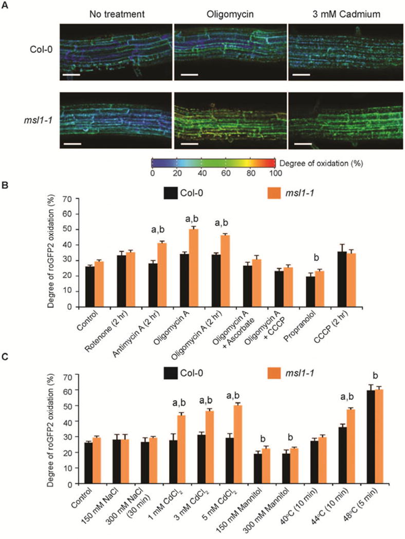
(A) Representative ratiometric pseudocolored images of mito-roGFP2 in Col-0 and msl1-1 roots treated with oligomycin and Cd2+. roGFP2 fluorescence was measured for excitation at 405 nm and 488 nm, and the degree of oxidation is calculated based on the ratio between the intensities from the two excitations (405/408 ratio) and the calibration of the probe using 10 mM DTT and 10 mM H2O2. The colour scale represents the redox state of roGFP2 ranging from fully reduced in indigo to fully oxidized in red. Scale bar = 50 μm. (B) Quantitation of mito-roGFP2 oxidation state in Col-0 and msl1-1 roots treated with respiratory inhibitors. Concentration of inhibitors used: 10 μM oligomycin A, 20 μM antimycin A, 20 μM rotenone, 1 mM sodium ascorbate, 1 mM propranolol 25 μM carbonyl cyanide m-chlorophenyl hydrazone (CCCP). (C) Quantitation of mito-roGFP2 oxidation state in Col-0 and msl1-1 roots under abiotic stress conditions. All treatments were carried out in the dark for 1 hr in ½MS medium (pH 5.7) unless otherwise stated. At least 10 seedlings in several independent experiments were examined, with error bars indicate standard error. “a” and “b” indicate significant difference (p < 0.01 based on ANOVA and Tukey post-hoc analysis), respectively, between Col-0 and msl1-1 under the same treatment and between control and treatment.