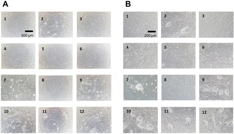
Figure 2.
The phase-contrast photomicrographs of iPS cells after cryopreservation (1)–(12). 1, ES cell culture medium; 2, ES cell culture medium containing 10% DMSO; 3, ES cell culture medium + 10% glycerol; 4, ES cell culture medium + 5% DMSO; 5, ES cell culture medium + 5% glycerol; 6, ES cell culture medium + 5% DMSO; 5% glycerol; 7, cell-freezing medium-DMSO; 8, cell-freezing medium-glycerol; 9, Cell Banker 1; 10, Cell Banker 1+; 11, Cell Banker 2; 12, Cell Banker 3. The photomicrographs were taken with ×40 (A) and ×100 (B) objectives. The iPS cells were cultured on mitomycin-treated MEF cells for 3 days after inoculation. Scale bars: 500 μm (A) and 200 μm (B).

