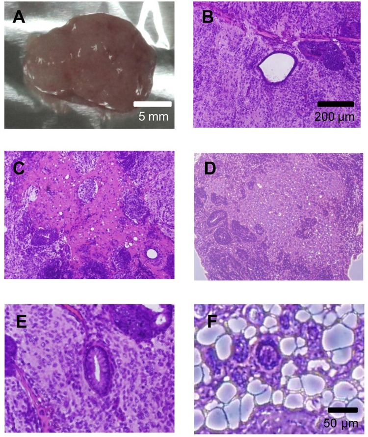
Figure 4.
Teratoma formation by iPS cells. Various tissues were present in teratomas derived from iPS cells. (A) Four weeks after the injection, the teratoma was surgically dissected from the mice. (B–F) Slides were stained with hematoxylin and eosin. (B) Artery-like structures. (C) Nerve-like structures. (D) Cartilage-like structures. (E) Gut epithelium-like structures. (F) Adipose-like structures.

