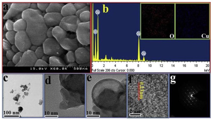
Figure 1.
Microscopic images of CuO NPs synthesized by a green method: (a) scanning electron microscopy (SEM) image; (b) EDAX spectrometry of the CuO NPs; inset: elemental mapping of oxygen and copper; (c–e) transmission electron microscopy (TEM) images at different magnifications; (f) high magnification view of the CuO NPs; and (g) Selected Area Electron Diffraction (SAED) pattern of the CuO NPs [39].

