Figure 1.
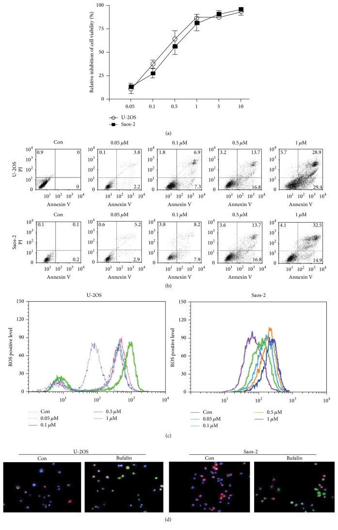
Bufalin regulates apoptosis, ROS production, and mitochondrial membrane potential in OS cells. (a) After treatment with various concentrations (0.05–10 μM) of bufalin for 24 h, viability of U-2OS and Saos-2 cells was determined using the CCK-8 assay. The inhibition ratio of cell viability increased in a dose-dependent manner. (b) U-2OS and Saos-2 cells were treated with 0.05–1 μM of bufalin and stained with Annexin V/PI. The apoptotic cells were detected by FCM. The apoptosis ratio changed in a dose-dependent manner. (c) After exposure to bufalin in different concentrations, fluorescence intensity indicating the intracellular ROS production was evaluated by FCM. ROS production increased gradually as bufalin concentration rose. (d) The ΔΨm of OS cells was measured using the JC-1 dye. After being treated with IC50 bufalin, the OS cells exhibited a reduction of ΔΨm as demonstrated by a decrease in red/green ratio. The red/green ratio in the control and bufalin group was 57.26 ± 7.13% versus 5.37 ± 0.63% for U-2OS cell, respectively (P < 0.01), while it was 60.63 ± 5.67% versus 3.83 ± 0.33% for Saos-2 cell (P < 0.01).

