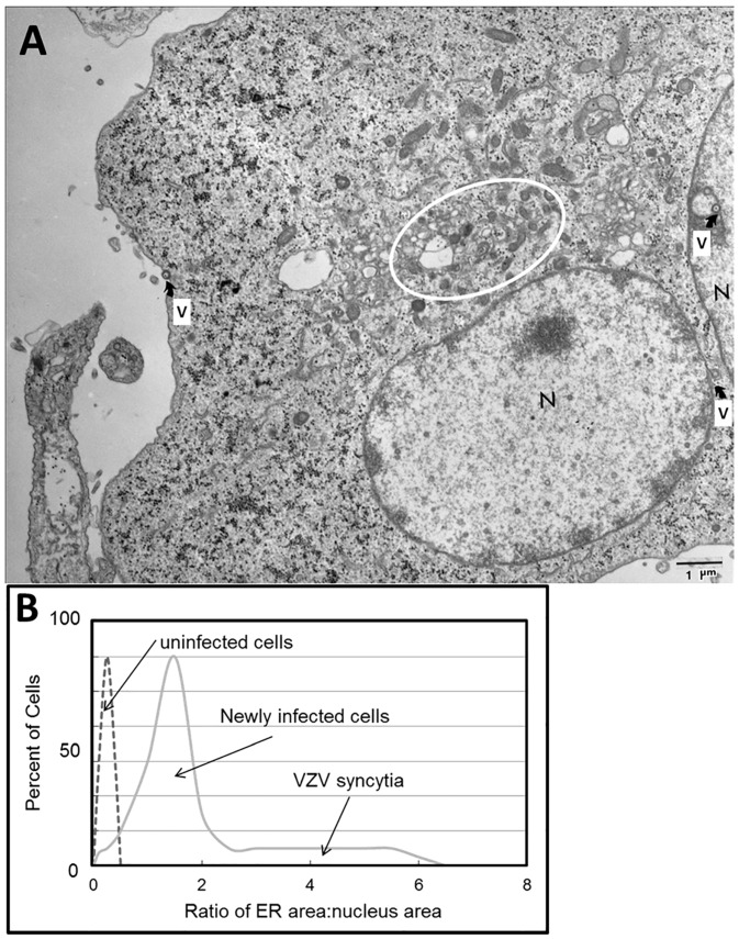
Figure 4.
Enlarged endoplasmic reticulum (ER) area in VZV-infected cells. (A) Electron micrograph of infected cells. The electron micrograph shows two adjacent VZV-infected cells that have fused. Each of two nuclei is marked by the letter N. Scattered viral particles are marked by the letter V with an arrow; these include capsids within nuclei and incompletely enveloped particles in the cytoplasm. The area surrounding the ER is encircled. (B) Ratio of ER area to nucleus area. Note the larger ratio as the degree of infection rises from newly infected cells to advanced infection with syncytia formation.

