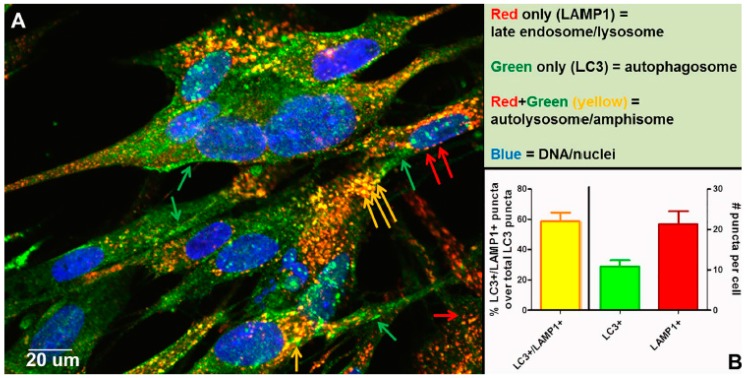
Figure 8.
Completion of autophagic flux during VZV infection. At 72 h post infection (hpi), infected cells were fixed with 2% paraformaldehyde and then processed for examination by confocal microscopy. Monolayers were incubated in primary antibodies: rabbit anti-LC3B and mouse anti-LAMP1. Monolayers were then incubated in the following antibodies: goat-anti-mouse-AlexaFluor 546, goat-anti-rabbit-AlexaFluor 488, as well as H33342. (A) Micrograph of a representative 400× image. Red arrows point to late endosomes/lysosomes (LAMP1+), green arrows point to autophagosomes (LC3+), and yellow arrows point to autolysosomes/amphisomes (LC3+/LAMP1+) (see Figure 7). (B) Quantitation of puncta. Puncta from many images were quantitated (>30 cells). The chart shows the percent puncta that were both LC3+ and LAMP1+ over total LC3+ puncta (left Y-axis), as well as average numbers of LC3+ puncta and LAMP1+ puncta per cell (right Y-axis).

