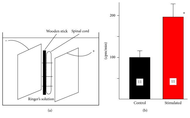
Figure 3.
The 45Ca2+ accumulation by the segments of the spinal cord stimulated in vitro by two electrodes of the same size. (a) An experimental setup. Two stainless steel plates (30 × 7 mm) were placed 9 mm apart inside of the plastic tube and connected to the source of DC. The segment of the spinal cord, attached to the wooden stick, was placed between two electrodes. (b) Accumulation of 45Ca2+ in the segment of the spinal cord (in cpm/mm) following 3 mA, 3′ stimulation. The increase in stimulated spinal cord (190.9%) is statistically significant (∗ p < 0.029, t-test).

