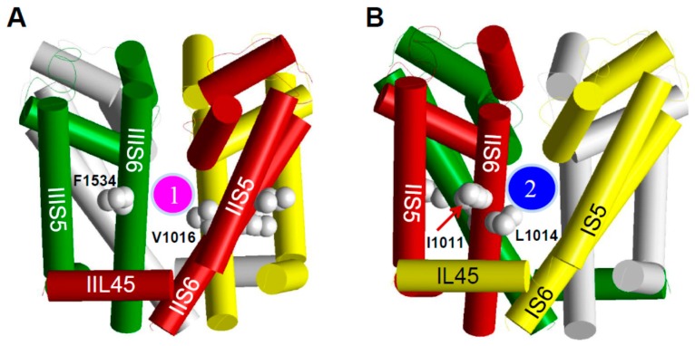
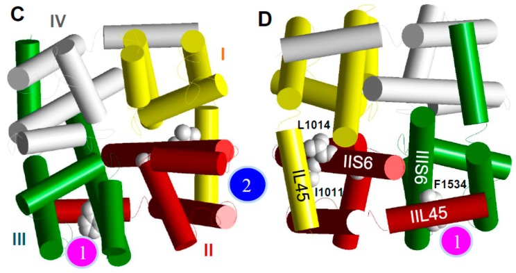
Figure 3.
The pore-domain model of the mosquito sodium channel. (A, B), Side views; (C) Extracellular view; (D) Intracellular view. Helical segments of the channel protein in domains I, II, III, and IV are shown as yellow, red, green, and gray cylinders, respectively. Location of the pyrethroid receptor sites PyR1 and PyR2 is indicated by the magenta and blue circles, respectively. The four positions where three kdr mutations are detected in the mosquito sodium channel and another kdr mutation (L1014F) in other insect sodium channels are shown as space-filled side chains of the wild-type residues. Only carbon atoms (gray spheres) in these side chains are shown, whereas the hydrogen atoms are removed for clarity. Note that opposite faces of helix IIS6 contain residues that contribute to PyR1 (V1016) or PyR2 (I1011 and L1014).


