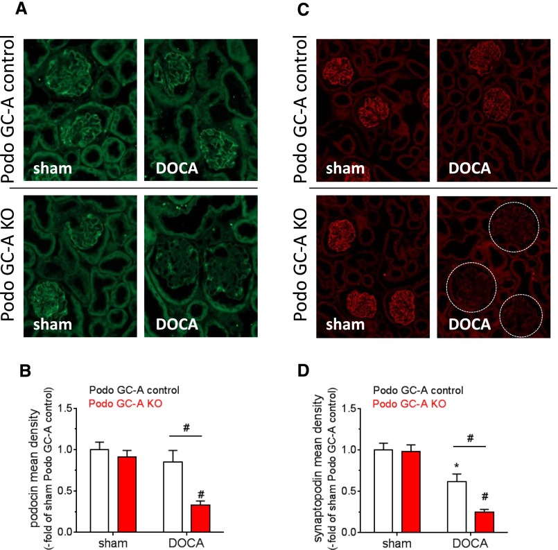
Figure 7.
The expression levels of the podocyte proteins podocin and synaptopodin are markedly reduced in Podo GC-A KO mice treated with DOCA. Immunofluorescence staining for podocin (A) and synaptopodin (C) showed a typical podocyte pattern in sham and DOCA-treated Podo GC-A control mice and in sham Podo GC-A KO mice. In contrast, podocin staining was restricted to podocyte cell bodies, and synaptopodin staining was hardly visible in DOCA-treated Podo GC-A KO mice. Analysis of fluorescence density (product of intensity and area) in glomeruli revealed significant downregulation of podocin expression in Podo GC-A KO mice by DOCA treatment (B). Glomerular synaptopodin density was reduced by DOCA in both genotypes, although to lower levels in Podo GC-A KO compared with Podo GC-A control (D). #P<0.001 versus sham or versus other genotype at same treatment as indicated. *P<0.05 versus sham.

