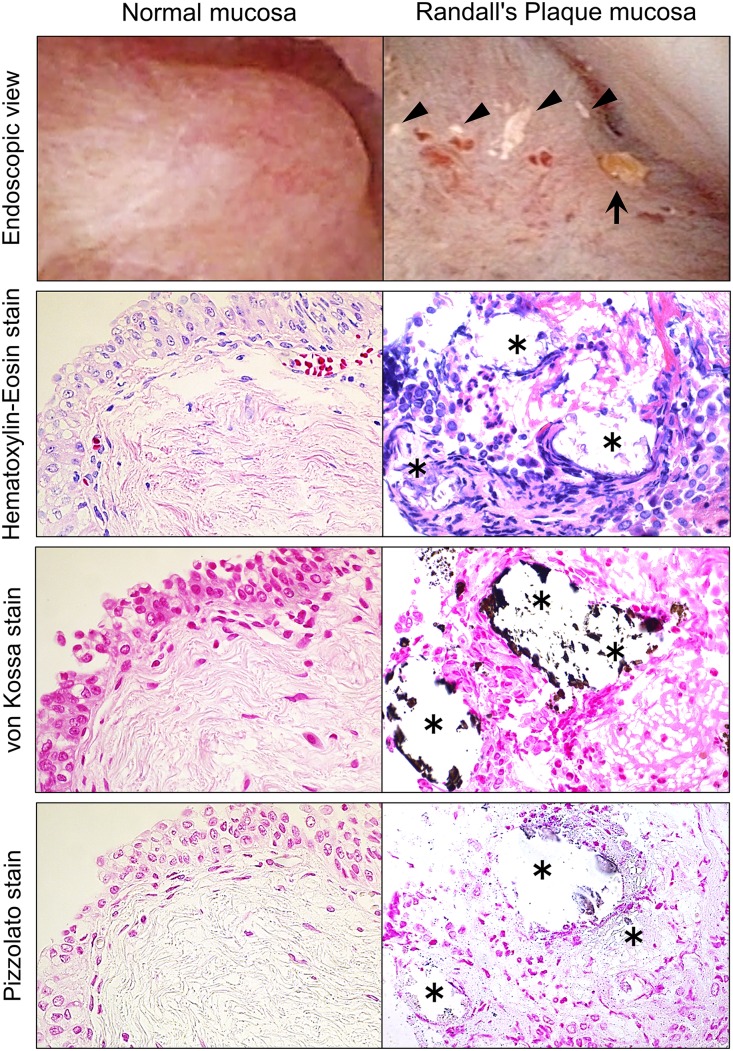
Figure 1.
Endoscopic and microscopic distribution of RPs. Representative photographs show renal papillary tissues from both normal and RP mucosa. The endoscopic image shows renal papilla mucosa in the upper calyx during retrograde intrarenal surgery. The normal papilla shows fleshy smooth mucosa without bleeding or calcification. Some RPs are showing as a white patchy lesion (arrow heads) as well as a ductal plug (arrow) within the same papilla. Micro tissues were stained with hematoxylin-eosin, von Kossa (for detection of CaP crystals), and Pizzolato (for detection of CaOx crystals) staining. *Location of RP. Magnification, ×400.

