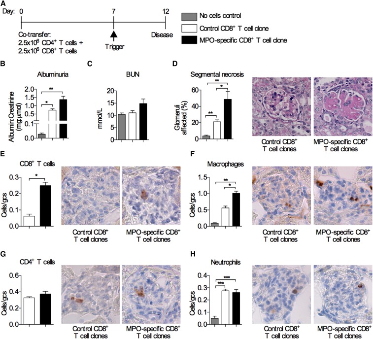
Figure 3.
Cotransfer of MPO–specific CD8+ T cell clones together with MPO–specific CD4+ T cells exacerbates experimental anti–MPO GN. (A) Timeline of MPO–specific CD4+ and CD8+ T cell transfer into Rag1−/− mice and the disease model. Control mice received MPO–specific CD4+ T cells and CD8+ T cell clones specific for OVA 257SIINFEKL264. Another control group received anti-GBM globulin alone without cells. Disease parameters assessed were (B) albuminuria; (C) BUN; (D) glomerular segmental necrosis (periodic acid–Schiff–stained micrographs show segmental necrosis in mice that received the MPO–specific CD8+ T cell clones); and infiltration of (E) CD8+ T cells, (F) macrophages, (G) CD4+ T cells, and (H) neutrophils (DAB Brown–stained micrographs depict illustrative examples of glomerular infiltration of the respective cell type). All bar graphs represent means±SEMs of n=4–6 per group. gcs, Glomerular cross-section. *P<0.05 by ANOVA; **P<0.01 by ANOVA; ***P<0.001 by ANOVA.

