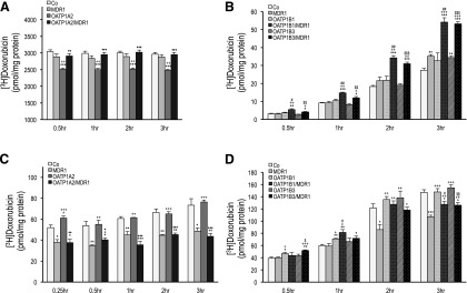
Fig. 5.
Vectorial transcellular transport and intracellular accumulation of doxorubicin. Radiolabeled doxorubicin (1.0 µM for OATP1A2; 0.2 µM for OATP1Bs) was administered to the apical (for OATP1A2) and basal (for OATP1Bs) compartment of monolayers of MDCKII-control (Co), single (MDR1, OATP1A2, OATP1B1, and OATP1B3)- and double (OATP1A2/MDR1, OATP1B1/MDR1, and OATP1B3/MDR1)-transfected cell lines. After incubation at given time points, translocation of doxorubicin into the apical compartment (A and B) and intracellular doxorubicin accumulation (C and D) are shown. Apical doxorubicin was significantly lower in MDCKII-OATP1A2 cells compared with control at all time points (A). Apical doxorubicin was significantly higher in double-transfected MDCKII-OATP1B1/MDR1 and MDCKII-OATP1B3/MDR1 cells compared with control at all time points (B). Intracellular doxorubicin was significantly higher in MDCKII-OATP1A2 (C) and MDCKII-OATP1B1 (D) cells at early time points. Data are expressed as mean ± S.E. (n = 4 for both studies). *P < 0.05, **P < 0.01, and ***P < 0.001 versus MDCKII-Co; +P < 0.05, ++P < 0.01, and +++P < 0.001 versus MDCKII-MDR1; ♦P < 0.05, ♦♦P < 0.01, and ♦♦♦P < 0.001 versus MDCKII-OATP1A2; #P < 0.05 and ##P < 0.01 versus MDCKII-OATP1B1; $P < 0.05, $$P < 0.01, and $$$P < 0.001 versus MDCKII-OATP1B3 cells.

