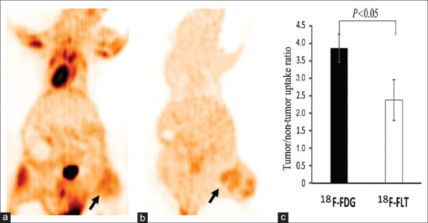
Figure 2.
Lung cancer xenograft images acquired by micro-PET. (a) 18F-FDG PET imaging of lung cancer animal xenograft. (b) 18F-FLT PET imaging of lung cancer animal xenograft. An subcutaneous cancer nodule of the left lower limb shows a mild heterogeneity in 18F-FDG uptake and 18F-FLT uptake (arrows). The tumor uptake degree of 18F-FDG PET was slightly higher than that of 18F-FLT. (c) The tumor-to-nontumor uptake ratio of the region of interest in animal xenografts was compared between the two tracers, using lung tissue as background (n = 6 per group). PET: Positron emission tomography; 18F-FDG: 2-[18F]-fluoro-2-deoxy-D-glucose; 18F-FLT: 3-deoxy-3-[18F]-fluorothymidine.

