
Figure 4.
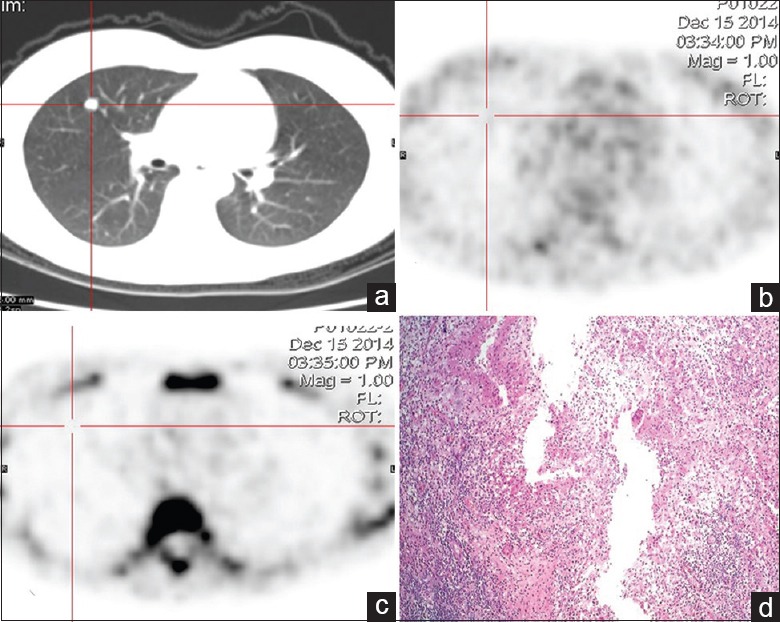
Dual-tracer PET/CT and histological images of a 43-year-old female with solitary pulmonary nodule in the right upper lung. (a) CT shows a small nodular opacity with a well-defined edge on the right upper lung. (b) 18F-FDG image shows no radioactivity uptake in the lesion. (c) 18F-FLT image also shows no radioactivity uptake in the lesion. (d) Result of pathological examination shows lung tuberculosis (HE, original magnification ×100). PET/CT: Positron emission tomography/computed tomography; 18F-FDG: 2-[18F]-fluoro-2-deoxy-D-glucose; 18F-FLT: 3-deoxy-3-[18F]-fluorothymidine.