Figure 2.
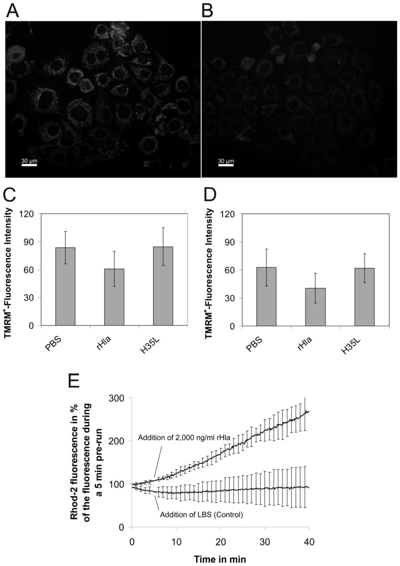
Exposure of airway epithelial cells to S. aureus α-toxin results in the reduction of inner mitochondrial membrane potential and calcium uptake into the matrix. 16HBE14o- or S9 cells were loaded for 1 h with 5 nmol/L of the fluorescent indicator dye tetramethylrhodamine methyl ester (TMRM+), which shows different subcellular localization and fluorescence intensities with changing values of the inner mitochondrial membrane potential. (A,B) examples of fluorescent images obtained in TMRM+-loaded 16HBE14o- cells kept under control conditions for 1 h (A) or after exposure to 2000 ng/mL rHla for 1 h (B); (C,D) TMRM+ fluorescence intensities in 16HBE14o- cells (C) or S9 cells (D) measured after 1 h of cell exposure to the vehicle (PBS, control), 2000 ng/mL rHla or rHla-H35L, respectively. Data are presented as means ± standard deviation (S.D.) (n = 4). Testing the data series for acceptance of the H0 hypothesis (no differences of means of all data series) using ANOVA revealed that this hypothesis had to be declined (p < 0.05) for 16HBE14o- cells (Figure 2C) and accepted for S9 cells (Figure 2D, p > 0.05). Comparisons of individual means (experimental vs. PBS controls) of data obtained using 16HBE14o- cells, however, did not reveal any significant differences; (E) Rhod 2-fluorescence indicating concentrations of free calcium ions in the mitochrondrial matrix of suspended and dye-loaded 16HBE14o- cells in the absence (control) or presence of 2000 ng/mL rHla (addition of vehicle or rHla at 5 min). Data are presented as means ± S.D. (n = 3).

