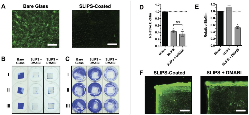
Figure 5.
SLIPS loaded with DMABI resist fouling on the substrate surface and inhibit biofilm formation on surrounding uncoated surfaces. Glass or SLIPS-coated substrates were submerged in P. aeruginosa cultures at the bottoms of the wells of 12-well microtiter plates and incubated for 24 h. Substrates were then removed and the attached biofilms were stained with either SYTO 9 or CV. (A) Representative fluorescence microscopy images of P. aeruginosa biofilm near the center of glass (left) or SLIPS-coated (right) substrates. (B) Representative image of CV-stained biofilms attached to glass substrates, SLIPS without DMABI, or SLIPS loaded with DMABI. (C) Representative image of CV staining of the bottoms of the wells of the 12-well microtiter plate (after the removal of the substrates shown in panel A). (D) Quantification of biofilm formed on the glass and SLIPS substrates shown in panel B. (E) Quantification of biofilm formed on the surrounding well bottoms shown in panel C, showing a reduction of biofilm in wells that contained DMABI-loaded SLIPS. (F) Representative fluorescence microscopy images of biofilm formed on the exposed glass edges of SLIPS substrates without loaded DMABI (left) and with loaded DMABI (right). Scale bars in A and F are 400 µm. Error bars in D and E represent the standard error of three independent experiments (n = 3). * = p <0.05. NS = no statistical difference.

