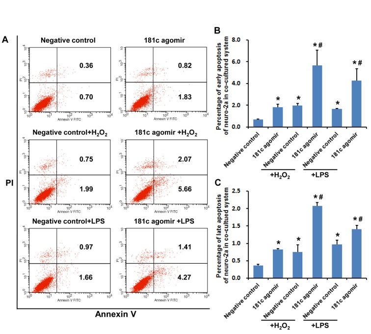
Figure 5.
MiR-181c agomir induces apoptosis of Neuro-2a cells co-cultured with BV2 microglial cells upon oxidative stress and inflammation. A) Apoptotic cells were detected by flow cytometry. Second and fourth quadrants represent late and early apoptotic fractions, respectively. B) Early apoptosis of Neuro-2a cells. C) Late apoptosis of Neuro-2a cells. *P < 0.05 vs. negative control group; #P < 0.05 vs. negative control + H2O2 or negative control + LPS group.

