
Fig. 1.
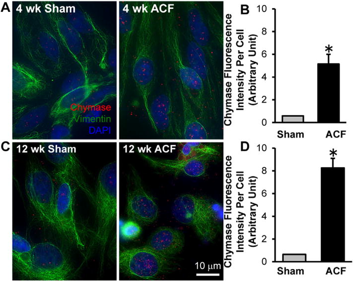
Chymase protein is increased in cardiac fibroblasts isolated from 4 and 12 week ACF rats compared to age-matched shams as shown by Immunocytochemistry. Cardiac fibroblasts were isolated from 4 week (A) and 12 week (C) sham and ACF rats followed by indirect fluorescence microcopy using chymase (red) and vimentin (green) antibodies. DAPI: blue. Quantitation of chymase fluorescence intensity in at least 40 cells demonstrates a marked increase in chymase protein in fibroblasts isolated from 4 week (B) and 12 week (D) ACF rats compared to the age-matched shams.