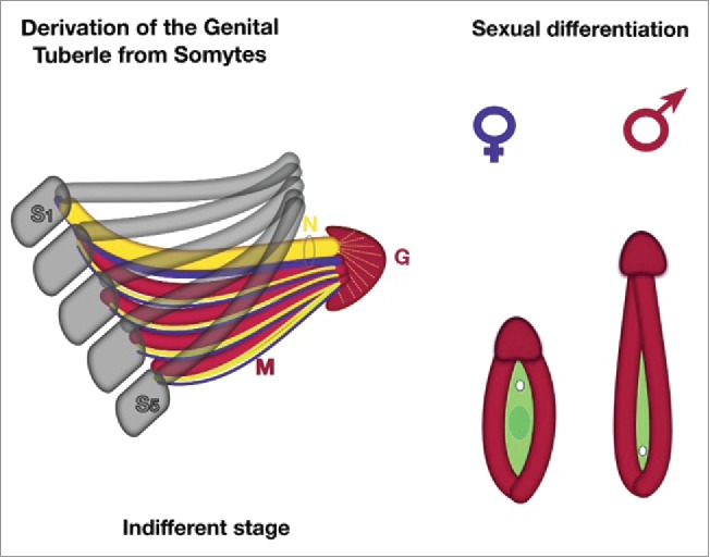
FIGURE 5.

New theory about derivation of external genitalia (schematic). On the indifferent stages the 5 sacral somites (S1-S5) have to recede of their segmentation and desintegrate. The sclerotomes (gray color) fuse to pelvic bones, which form the arcus, they conjoin together end-to-end in the midline of ventral body with pubic symphysis. The fused 5 sacral myotomes (M, red color) with its genuine neurotomes (blue) and angiotomes (yellow), covered by dermatome growing below along of pubic bones and fuse together endways on pubic area. The endwise conjoined myotomes form the corpora cavernosa of genital tubercle, following with dorsal neuro-vascular bundles (bold yellow and blue in the ring, labeled N). The top of myotomes ends become glans tubercle (G) covered of ectodermal layer. During sexual differentiation: myotomes are forming the genital swelling (red), genital tubercle become glans penis or clitoridis, urogenital fold (green) originating from cloaca (hindgut) of embryo. Female pattern or indifferent stage – small tubercle with opened vestibulum vaginae. The genital swellings form the labial folds; the branches of the genital tubercle (corpora cavernosa) surround the urogenital groove and located deep inside in the genital swellings. Male pattern – fusion of labial folds into the urogenital sinus (or penile urethra) extends along the elongated phallus. The branches of the genital tubercle (corpora cavernosa) conjoin together.
