
FIGURE 5.
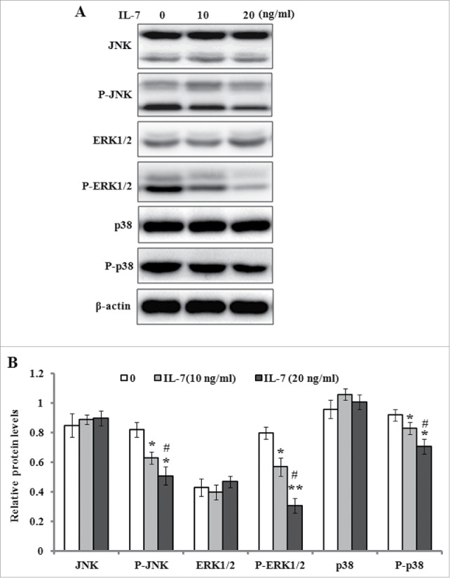
IL-7 inhibits the MAPK signaling in PDLSCs. PDLSCs were stimulated with IL-7 (0, 10 and 20 ng/ml) for 48 h, and total protein was extracted and subjected to Western blot analysis. (A) Representative western blot images demonstrating a markedly inhibitory role on the phosphorylation levels of JNK, ERK1/2 and p38, but not their protein levels. (B) Relative protein levels of JNK, ERK1/2, p38, and their phosphorylated levels were quantified by the densitometry of each band normalized to β-actin signal. Data are presented as mean ± SD, *P < 0.05 and **P < 0.01 compared with the control (0 ng/ml of IL-7). #P < 0.05 compared with the group (10 ng/ml of IL-7).