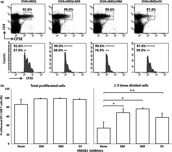
Figure 3.

Carboxyfluorescein succinimidyl ester (CFSE)‐labeled OT‐1 cell‐transfused B6 mice were immunized by s.c. injection of OVA 257–264 peptide (OVA) with topical imiquimod (IMQ) and high mobility group box 1 (HMGB1) inhibitors (200 μg/mouse) (four mice/group), and the proliferation of OT‐1 CD8 T cells was examined 3 days after the immunization by FACS analyses. (a) Representative staining patterns of each treatment group are shown. The boxes and the upper bar (histograms) indicate the proliferated OT‐I CD8 T cells. Lower bars in the histograms indicate the OT‐I CD8 T cells that were divided at least three times. (b) Contents of total OT‐I CD8 T cells (left) and OT‐I CD8 T cells divided at least three times (were) are shown. Data are representative of two experiments. *P < 0.05 by anova. GM, gabexate mesilate; IMQ, imiquimod; NM, nafamostat mesilate; SV, sivelestat.
