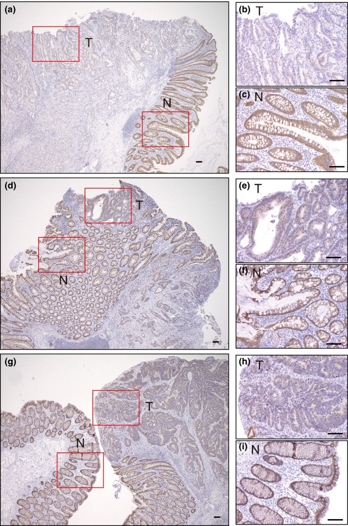
Figure 3.

Immunohistochemical staining of ABCC3 in colon cancer and normal colon tissue. Tissue sections containing normal (N) and tumor (T) cells are from patients 1 (a–c), 2 (d–f), and 3 (g–i). Boxed regions in (a), (d), and (g) are shown at higher magnification in (b) and (c), (e) and (f), and (h) and (i), respectively. Scale bars, 100 μm.
