
Figure 4.
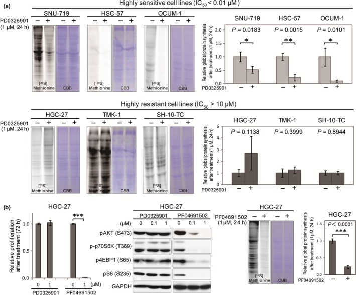
Suppression of global protein synthesis after MEK inhibition is involved in sensitivity to MEK inhibition. (a) Sensitive (SNU‐719, HSC‐57, and OCUM‐1) and resistant (HGC‐27, TMK‐1, and SH‐10‐TC) cell lines were treated with DMSO or 1 μM PD0325901 for 24 h and then subjected to [35S]methionine incorporation assay. Equal loading of the proteins was shown by staining with Coomassie brilliant blue. Intensities of protein bands labeled with [35S]methionine were quantified by ImageJ software, and normalized against those labeled with Coomassie brilliant blue. Representative band images are shown on the left. Differences in incorporated [35S]methionine between cells treated with DMSO (n = 3) and 1 μM PD0325901 (n = 3) were analyzed by Student's t‐test and shown on the right. (b) HGC‐27 cells were treated with DMSO, PD0325901, or PF04691502 (0.1 or 1.0 μM) and subjected to cell proliferation analyses and Western blotting with antibodies against pAKT, p‐p70S6K, p4EBP1, and GAPDH (left). HGC‐27 cells treated with DMSO or 1 μM PF042691502 were also subjected to [35S]methionine incorporation assay (right). *P < 0.05, **P < 0.01, ****P < 0.0001.