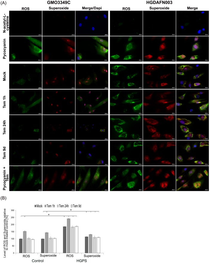
Fig 5. Temsirolimus does not impact the levels of reactive oxygen species and superoxide in HGPS cells.
(A) Immunochemistry was performed on control (GMO3349C) and HGPS (HGADFN003) fibroblasts mock-treated or temsirolimus-treated for the indicated periods. Live cells were stained with an oxidative stress detection reagent for ROS and a superoxide detection reagent. The negative control was treated with N-acetyl-L-cysteine to quench the signal intensity. The positive control was treated with pycocyanin to induce ROS production. DNA was stained with Hoechst 33342. Representative images are shown (n = 4). Scale-bar: 20 μm. (B) The same cells and detection reagents as in (A) were used to perform a 96-well microplate assay as described in Methods. The percentage of activity was calculated relative to mock-treated control cells. Data are expressed as the mean ± S.D. (*p-value ≤ 0.05; n = 4).

