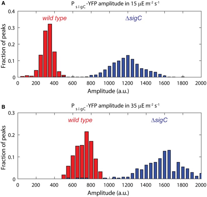
Figure EV2. PsigC‐YFP levels are upregulated in high light, suggesting a reduced functionality of SigC in higher light conditions.

- Histogram of PsigC‐YFP expression in wild‐type (red) and sigC deletion mutant (blue) under ca. 15 μE m−2 s−1 cool white light. For the wild type, 664 cells from nine movies were collected, whereas for the sigC deletion strain, 1,084 cells from six movies were collected.
- Histogram of PsigC‐YFP expression in wild‐type (red) and sigC deletion mutant (blue) under ca. 35 μE m−2 s−1 cool white light. PsigC‐YFP in wild type is upregulated by twofold between light conditions. A smaller fold change of 1.2 is seen in the sigC deletion mutant. In the higher light condition, 1,003 cells from two movies were collected for the wild‐type strain, whereas 920 cells from two movies were collected for the sigC deletion strain.
Source data are available online for this figure.
