
Fig. 3.
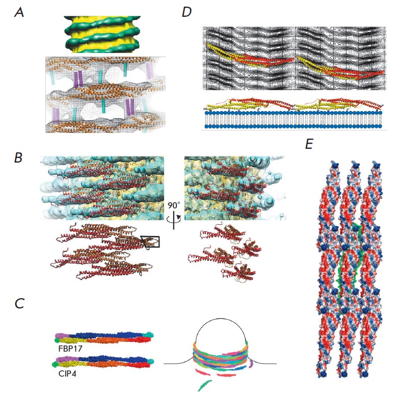
Oligomerization of BAR domains on membranes. A – The cryo-electron microscopy model of a 28-nm membrane tubule with endophilin oligomers (top image). The BAR domain (orange) and additional helices (cyan and magenta) are fitted into the electron density [7]. B – Oligomerized BAR domains of amphiphysin 2 [72]. C – Scheme of BAR domain oligomerization and formation of membrane tubules [16]. D – Interactions between dimerized BAR domains of CIP4 [73]. E – Interactions between dimerized BAR domains of Pinkbar [45].