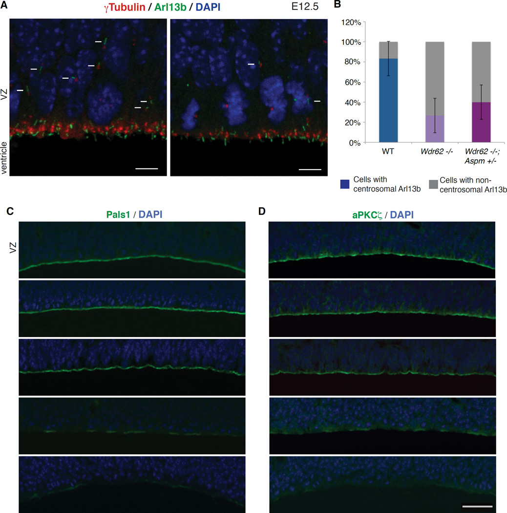
Figure 7. Loss of Wdr62 and Aspm disrupts apical complex proteins and leads to premature dissociation of ciliary remnants from centrosomes.
(A) Reduction in centrosomes and cilia in the Wdr62 −/− mouse brain, both at and away from the ventricular surface, along with an increase in dissociated ciliary remnants. Shown here are E12.5 WT (top) and Wdr62 −/− (bottom) brain sections immunostained for γ-tubulin (centrosomes) and Arl13b (cilia). Arrows denote cilia located away from the ventricular surface, while arrowheads mark non-centrosomal Arl13b (dissociated ciliary remnants). Scale bar = 5 µm.
(B) Quantification of centrosomal versus non-centrosomal Arl13b in WT, Wdr62 −/− and Wdr62 −/−; Aspm +/− brains. Fisher’s exact test, ** p = 0.002 for WT vs. Wdr62 −/− and ** p = 0.009 for WT vs. Wdr62 −/−; Aspm +/−.
(C–D) Loss-of-function of Wdr62, Aspm or both severely disrupts apical complex proteins Pals1 and aPKCζ in a dose-dependent manner. From top to bottom: WT, Wdr62 +/−, Aspm −/−, Wdr62 +/−; Aspm −/− and Wdr62 −/− brains at E14.5 stained for (C) Pals1 and (D) aPKCζ to label the apical complex, and counterstained with DAPI. Scale bar = 50 µm.

