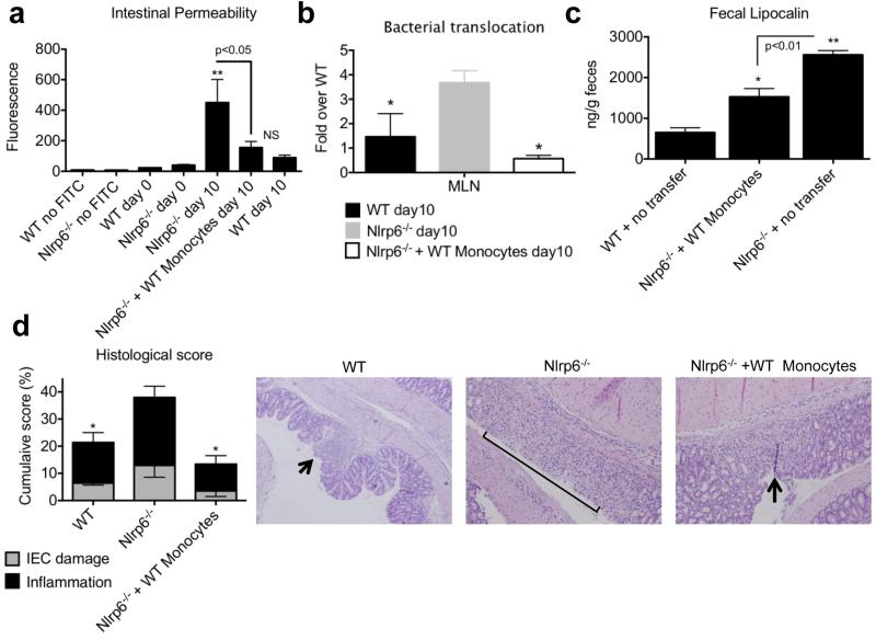
Figure 3. Adoptive transfer of WT Ly6Chi monocytes into Nlrp6−/− mice limits bacterial translocation and intestinal damage.
Age- and sex-matched Nlrp6−/− and WT mice were treated with AOM followed by 5 days of 2% DSS. WT Ly6Chi monocytes were adoptively transferred into Nlrp6−/− mice 3.5 days after start of DSS. (a) Mice were gavaged FITC-dextran at end of 5 days of DSS followed by serum collection and measurement of fluorescence 4 hours later. (b) Normalized levels of total bacteria/MLN as measured by qPCR after 5 days of 2% DSS. (c) Fecal lipocalin-2 levels as measured by ELISA after 5 days of DSS. (d) Histological inflammatory scores based on extent of inflammatory cell infiltration and IEC damage;Micrographs of H&E sections are at 200x magnification. Arrow points to focal erosion with inflammatory infiltrate. Bracket indicates ulcerated epithelium with inflammatory infiltrate in LP and submucosa. Data are representative of three independent experiments, n=15, n=16, n=20 for WT, Nlrp6−/− and Nlrp6−/− + WT Ly6Chi monocytes groups respectively. *, ** - p<0.05, p<0.001, respectively, as compared to day 0 of both genotypes (a), or as compared to Nlrp6−/− (b,c) or as compared to WT (d).

