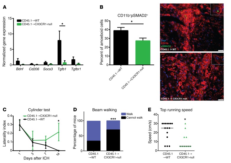
Figure 3. Microglial alternative activation is required for functional recovery after ICH.
(A) Microglia were cell sorted from WT and CX3CR1-null BM chimeras 14 days after ICH and then processed for qRT-PCR. CX3CR1-null microglia have a significant reduction in TGF-β1 gene expression compared with WT microglia. Means graphed with SEM; Student’s t test; n = 7–9. (B) Brain sections from WT and CX3CR1-null BM chimeras were stained for CD11b (red) and pSMAD2 (green) 14 days after ICH. Nuclei were stained with DAPI. ×40 images, with inset at ×64. Scale bars: 25 μm. Quantification of percent of pSMAD2+, amoeboid CD11b+ graphed as mean with SD; Student’s t test; n = 4. (C) BM chimeras were cylinder tested 1, 3, 7, and 14 days after ICH. CX3CR1-null chimeras maintain a right forelimb preference 14 days after ICH. Means graphed with SEM. Repeated measures ANOVA followed by Tukey’s post hoc test; n = 10–12. (D) Beam walking test shows that CX3CR1-null BM chimeras do not walk as well on the beam 14 days after ICH. Percentage of mice able versus unable to walk is graphed. Wilcoxon rank sum test; n = 11–13. (E) Forced run test shows that CX3CR1-null BM chimeras do not run as quickly as WT BM chimeras 14 days after ICH. Individual dots represent individual mice, with line at the median; Mann-Whitney U test, n = 10–15. *P < 0.05, ***P < 0.001.

