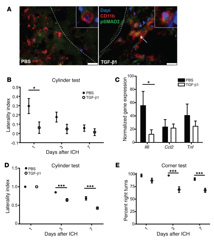
Figure 5. TGF-β1 treatment promotes better functional outcomes and modulates microglial IL-6 after ICH.
(A) TGF-β1 treatment led to phosphorylation of SMAD2 in myeloid cells in the brain. Brain sections from PBS- or TGF-β1–treated mice were stained at 24 hours for CD11b (red) and pSMAD2 (green). Nuclei were stained with DAPI. Arrow indicates cluster of pSMAD2+CD11b+ cells. Dotted line depicts edge of hematoma. 40×, with inset at 64×. Scale bars: 25 μm. n = 4–5. (B) Cylinder test shows that TGF-β1–treated mice have better functional outcomes 24 hours after ICH. Repeated measures ANOVA, followed by a Tukey’s post hoc test; n = 11–12. (C) TGF-β1–treated microglia have reduced Il6 gene expression 24 hours after ICH. Means graphed with SEM; Student’s t test; n = 7–9. (D and E) TGF-β1–mediated improvements in behavioral outcomes were confirmed using the collagenase model of ICH. Cylinder test (D) and corner test (E) show TGF-β1–treated mice have better functional outcomes at 3 and 7 days after ICH. Repeated measures ANOVA, followed by Tukey’s post hoc test; n = 9–10. *P < 0.05, ***P < 0.001.

