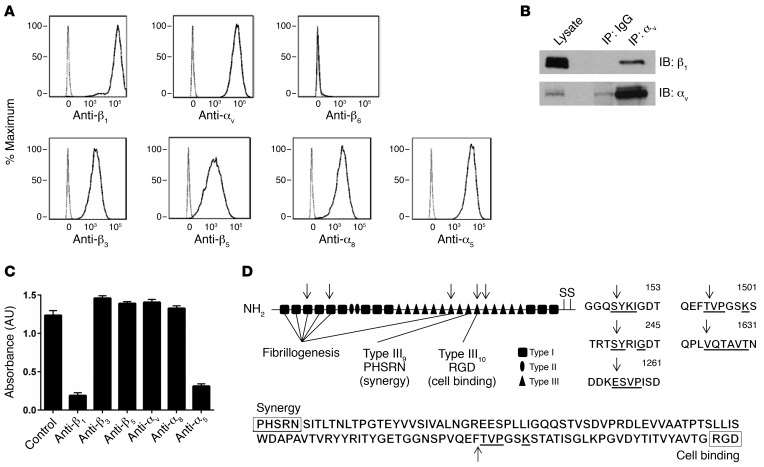
Figure 4. Integrin α5β1 is critical for smooth muscle cell adhesion to fibronectin.
(A) Human ASM cells in suspension were labeled with primary antibodies specific for cell-surface integrins β1, αv, β6, β3, β5, α8, α5 and a secondary antibody conjugated to APC. The cells were analyzed by flow cytometry and gated for live cells. The resultant population was analyzed for APC expression (solid line). Human ASM cells labeled with a secondary antibody alone served as a control (dashed line). Representative histograms of APC expression versus cell counts are shown. The x axis represents APC expression (mean fluorescence intensity); the y axis represents the cell count (percentage of maximum). (B) Representative Western blot for expression of integrin αvβ1 was determined by IP. Lysates from human ASM cells underwent pulldown with mouse IgG or anti-αv antibody, followed by immunoblotting (IB) for β1. Immunoblotting for αv was performed to confirm enrichment of αv. n = 3 distinct experiments. (C) Adhesion (measured by absorbance of crystal violet at 595 nm) of human ASM cells to fibronectin (0.1 μg/ml) after treatment with the indicated specific integrin-blocking antibodies. Data represent the mean ± SEM from triplicate experiments. (D) Schematic of fibronectin, with arrows marking chymase cleavage sites identified during N-terminal sequencing of the 3 cleaved fibronectin bands seen on the Coomassie staining in Figure 3A. The underlined amino acids correspond to aligned sequences detected during N-terminal sequencing.

