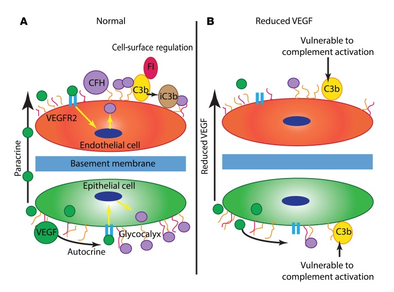
Figure 7. Proposed model of how VEGF regulates local complement activity.
(A) Under normal circumstances in the outer retina and renal glomerulus, specialized epithelial cells (the RPE and podocytes, respectively) produce VEGF that has both autocrine effects and paracrine effects on neighboring endothelial cells. VEGF signaling through VEGFR2 causes inhibitory complement proteins such as CFH to be produced by these cells. These complement inhibitors function at the cell surface to prevent complement activation caused by spontaneous alternative pathway hydrolysis. In the case of CFH, this occurs by binding to the cellular glycocalyx and acting as a cofactor for factor I (FI). (B) When anti-VEGF therapy is given, there is reduced local VEGF production, which leads to less VEGFR2 signaling. This may cause a reduction in local complement inhibitor synthesis and secretion, making the cells more vulnerable to complement activation.

