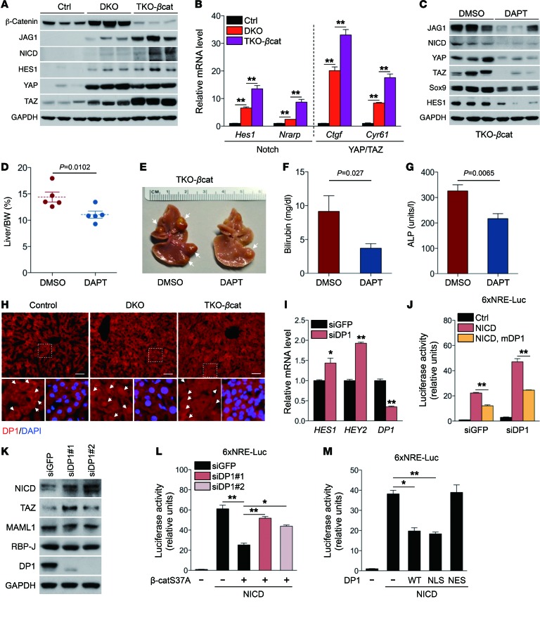
Figure 5. Wnt/β-catenin signaling inhibits YAP/TAZ/Notch positive feedback activation by promoting DP1 nuclear localization.
(A) Western blot analysis of control, DKO, and TKO-βcat liver tissue for the indicated proteins. (B) qRT-PCR analysis of Notch and YAP/TAZ target gene expression in liver from mice of the indicated genotypes (n = 3 per group). (C) TKO-βcat mice were given 20 i.p. injections of DAPT at a concentration of 125 mg/kg every other day. Western blot analysis of liver lysates from DAPT- or DMSO-injected TKO-βcat mice. (D–G) Analysis of liver-to-BW ratio (D), liver tumor size (E), serum levels of bilirubin (F) and ALP (G) in DMSO- or DAPT-injected TKO-βcat mice (n = 5 per group). (H) DP1 staining was performed on liver sections from 6-week-old mice of the indicated genotypes. Scale bars: 50 μm. Original magnification: ×25. (I) qRT-PCR analysis of Notch target gene expression in DP1-depleted cells (n = 3). (J) Notch-dependent reporter assay in Huh7 cell–depleted human DP1. The effect of DP1 loss on Notch reporter activity was rescued by overexpression of siRNA-resistant murine DP1 (mDP1) (n = 3). (K) Western blot analysis with the indicated antibodies in DP1-depleted Huh7 cells. (L) Notch reporter assay in Huh7 cells transfected with the indicated plasmids and siDP1 (n = 3). (M) Suppression of NICD-mediated reporter activity by WT DP1 or NLS-DP1, but not by NES-DP1 (n = 3). Data are expressed as the mean ± SEM. *P < 0.05 and **P < 0.01, by 2-tailed Student’s t test (F, G, and I) and 1-way ANOVA with Tukey’s post-hoc test (B, J, L, and M) when ANOVA was significant.

