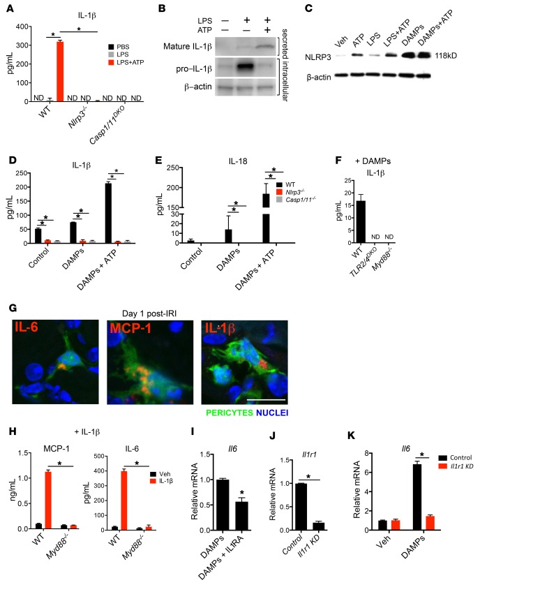
Figure 2. Activation of NLRP3 inflammasome by pericytes in response to DAMPs.
(A) Concentration of secreted IL-1β in WT and Nlrp3–/– and Casp1/11DKO pericytes 24 hours after treatment with LPS and ATP. (B) Western blot of intracellular pro–IL-1β and secreted mature IL-1β from pericytes 24 hours after treatment with LPS and ATP. (C) Western blot of pericyte cell lysates 24 hours after treatment with LPS, ATP, and DAMPs. (D and E) IL-1β and IL-18 concentration in WT and mutant pericyte supernatants after treatment with DAMPs and ATP. (F) IL-1β concentration in supernatants of WT, TLR2/4DKO, and Myd88–/– pericytes 24 hours after treatment with DAMPs. (G) Immunofluorescence staining for IL-1β, IL-6, and MCP-1 on sections from kidneys on day 1 after IRI. (H) IL-6 and MCP-1 cytokine response to IL-1β treatment in the supernatant of WT and Myd88–/– pericytes. (I) The effect of recombinant IL-1 receptor antagonist (IL1RA) on transcriptional response to DAMPs. (J and K) The effect of siRNA against Il1r1 on Il1r1 expression (J) and Il6 response to DAMPs (K). ND, not detected. KD, knockdown. (Scale bar: 25 μm; n = 3–6 per group; *P < 0.05, 2-tailed Student’s t test or 2-way ANOVA, Bonferroni’s multiple comparisons test.)

