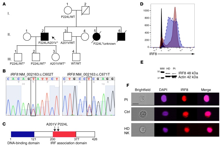
Figure 1. Linkage analysis of IRF8 compound heterozygous mutations in familial NKD and genetic confirmation of mutation.
(A) Pedigree of affected family. Roman numerals indicate generations. Arrow points to proband. Shown are amino acid variants in IRF8 as detected by whole exome sequencing. Asterisk indicates variant confirmed by Sanger sequencing. (B) Confirmation of proband’s IRF8 variants by Sanger sequencing. (C) Schematic of human IRF8 showing affected residues found within the IRF association domain. (D) Protein expression of IRF8 in EBV-transformed patient B cell line (maroon, solid line) relative to normal control (blue, dashed line). Isotype control is shown in black. Shown is 1 representative experiment from 3 independent repeats. (E) Western blot of IRF8 (48 kDa) in healthy donor (HD) or patient (Pt) lymphoblastoid cell lines with actin (42 kDa) as a loading control. Shown is 1 representative experiment from 3 independent repeats. (F) Nuclear localization of IRF8 in patient (Pt) or control (Ctrl) B cell lines or healthy donor NK cells. Cells were fixed and stained for IRF8 (eFluor 710, red) followed by counterstaining with DAPI (blue). Scale bar: 10 μm. Shown is 1 representative experiment from 3 independent repeats.

