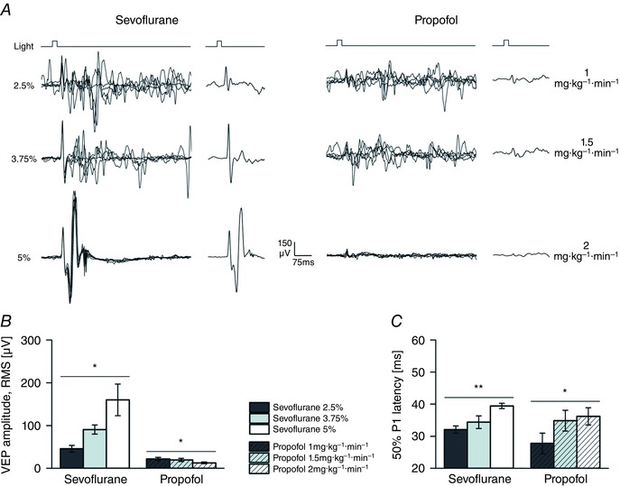
Figure 3. Opposite changes induced by sevoflurane and propofol on visual response.

A, single VEP traces and ensemble averages from an individual animal exposed to brief light pulses (pulse duration 20 ms; pulse irradiance 25 μW cm−2; pulse rate 0.1 Hz) at incremental concentrations of sevoflurane (2.5, 3.75 and 5%; left) and propofol (1, 1.5 and 2 mg kg−1 min−1; right). B and C, bars graphs of VEP amplitude (B; RMS amplitude, first 150 ms from stimulus onset, estimated from the averaged waveform) and the onset of the P1 deflection (C; mean latency of the 50% of P1 amplitude). These estimates were obtained from the same animals, each exposed to all dosages of both anaesthetics in two separate experimental sessions, run a few days apart (n = 6 rats). Notice how, sevoflurane increased the VEP amplitude in a concentration‐dependent fashion, while propofol did the opposite. Also notice that the VEP latency was incremented by both drugs, indicating a common reduction in neuronal excitability. Asterisks indicate significance values (B and C, principal effect by one‐way rANOVA; P > 0.05, ns; * P < 0.05; ** P < 0.01; *** P < 0.001).
