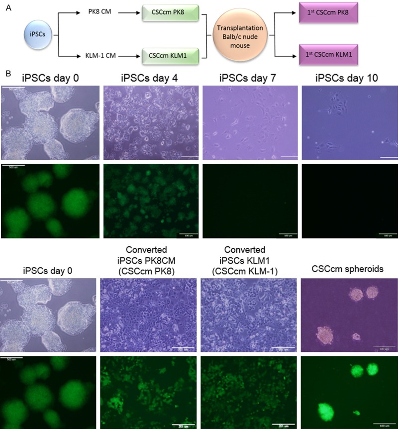
Figure 1.

Conversion. A. Representative scheme of the conversion procedure. B. Viability of iPSCs maintained in control medium without LIF was no longer than 10 days. iPSCs underwent differentiated with no remaining GFP positive cells and eventually expired (upper panel). Stemness tracking during conversion by the presence of GFP protein. Self-renewing potential was validated by sphere formation assay previously to the subcutaneous implantation (lower panel). Original magnification 4× and 20×.
