Figure 2.

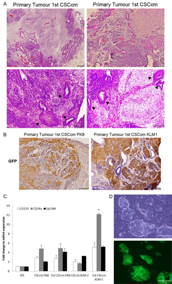
Malignant transformation and CSCs features. A. Histopathological features of 1st CSCcm primary tumours were evaluated by H&E staining. Specific PanIN lesions are indicated with arrowheads. Original magnification 10× and 20×. B. Lineage tracing by GFP protein showed that it was predominantly expressed in undifferentiated cells, however was also partially found in ductal-like structures. Original Magnification 10× and 20×. C. RT-qPCR analysis of CSCs markers CD133, CD24a and EpCAM in converted cells CSCcm and primary cultures 1st CSCcm. D. Cell distribution of primary cultures (1st CSCcm) after puromycin enrichment.
