
Figure 6.
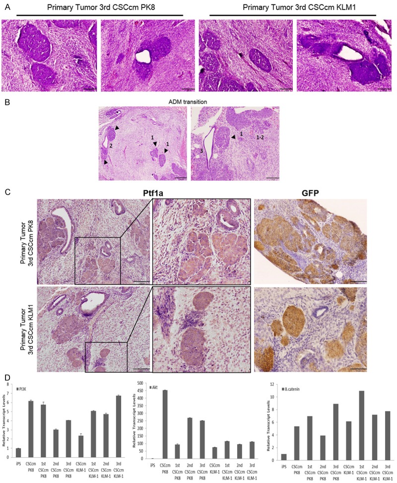
Acino-ductal metaplasia. A. H&E staining of cluster cells areas with acinar morphology and ADM structures found in 3rd CSCcm primary tumours. B. Images showing evidences of ADM transition originated from acinar structures (arrowheads) indicated as 1, acino-ductal interphase as 2 and final ductal-like acquired phenotype 3. C. Cell clusters and few ductal cells were positive for the acinar marker Ptf1a. GFP was equally predominantly located in cell clusters and few cells from ductal epithelial cells. D. RT-qPCR of the relative transcript levels for PI3K, AKT and β-catenin (from left to right) in a panel of the samples obtained from serial transplantation. Micrographs original magnification 10× and 20×.