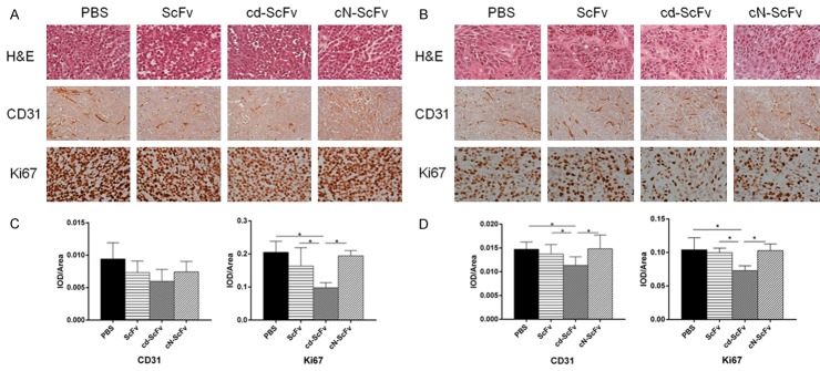
Figure 6.
Representative images of H&E staining and Immunohistochemistry of xenografted tumor. (A, B) Histological features of tumor specimens and representative photomicrographs of anti-CD31 and anti-Ki67 immunostaining on A549 (A) and L78 (B) xenograft after the mice were treated with ScFv, cdGIGPQc-ScFv, cNAQAEQc-ScFv for 2 weeks. Scale bar: 100 μm. (C, D) The IOD/Area of anti-CD31 and anti-Ki67 positivity in images of A549 (C) and L78 (D) xenograft was analyzed and calculated with Image-Pro Plus analysis software. At least 9 images from each group were used for quantification and statistical analysis. Statistical analyses were performed using unpaired t test. *, P<0.05.

