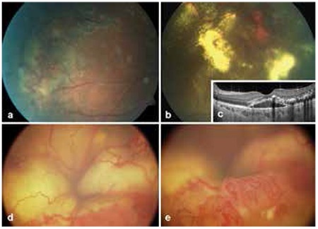
Figure 1. Images from various stages of Coats’ disease, a) peripheral telangiectatic vessels and exudation in stage 2A disease, b) telangiectatic vessels and exudation in stage 2B, c) optical coherence tomography image from the patient in Figure 1b showing subretinal fluid and hyperreflective exudation at the macula, d) image from a patient referred for intraocular tumor, stage 4 disease with total retinal detachment, e) Inferior peripheral telangiectatic vessels of patient in Figure 1d.

