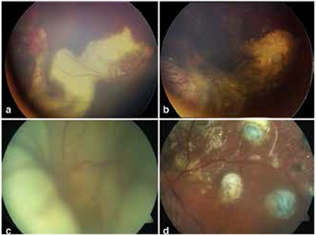
Figure 3. Fundus photographs taken before and after treatment, a) Telangiectatic vessels in the temporal periphery and exudation extending to the macula in a patient with stage 2B Coats’ disease, b) Post-treatment image from the same patient showing cryo scars, fibrotic nodule at the macula and exudation following cryotherapy, c) Pre-treatment image from a patient with total retinal detachment, d) Image from the same patient following pars plana vitrectomy, internal drainage, endophotocoagulation and endocryotherapy.

