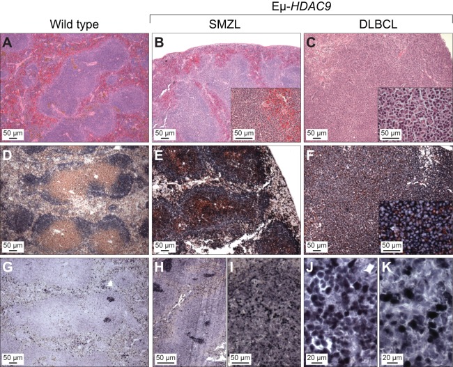
Fig. 4.
Histopathological analysis of mature B-cell lymphomas in Eµ-HDAC9 mice. Representative images showing disorganization of lymphoid tissue and expression of B220, CD3 and BCL6. Mouse spleen sections from wild type, splenic marginal zone lymphoma (SMZL) and diffuse large B-cell lymphoma (DLBCL) cases (as indicated) were stained with hematoxylin-eosin (A-C), doubled stained with B220 (blue)/CD3 (brown) (D-F), or BCL6 (blue) (G-J). IRF4 expression is shown in panels I and K.

