Fig. 2.
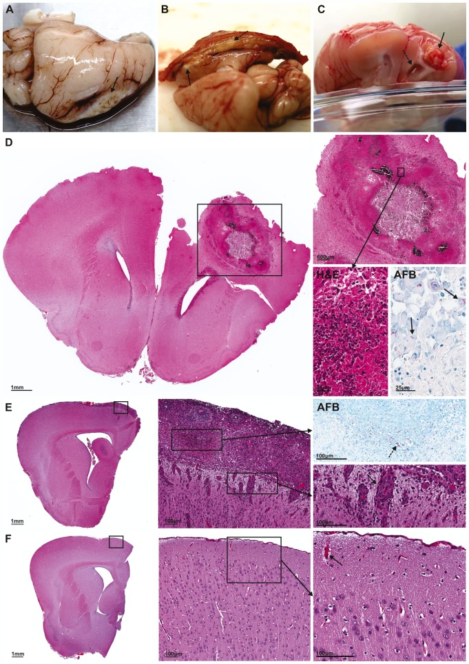
Representative gross pathological and microscopic examination of brain lesions. (A) Superficial, medial tuberculoma formed at 14 days post-infection. (B) Medial tuberculoma and exudate (dashed arrow) adhering brain to skull at 21 days post-infection. (C) Deep tuberculoma associated with hydrocephalus (dashed arrow) formed bilaterally but greater on side ipsilateral to tuberculoma at 35 days post-infection. Tuberculomas are indicated by small black arrows in panels A-C. (D) Microscopic examination of tuberculoma from panel C demonstrates the localization of the tuberculoma to one hemisphere. High-power view (inset) demonstrates central necrosis and dense cellular rim (H&E), with Ziehl–Neelsen stain (AFB) highlighting the numerous M. tuberculosis bacilli in the cellular rim. M. tuberculosis bacilli stained as red rods (black arrows). (E) Microscopic examination of one hemisphere of M. tuberculosis-infected brain with inflammatory meningitis. High-power view (inset) highlights numerous red M. tuberculosis bacilli (dashed arrow) with Ziehl–Neelsen stain (AFB) and demonstrates perivascular infiltrate (black arrow) associated with the inflammatory exudate (H&E). (F) Microscopic examination of one hemisphere of control brain. High-power view (inset) demonstrates no inflammatory exudate and normal vessels (black arrow) with no perivascular infiltrate.

